Nanotechnology Images
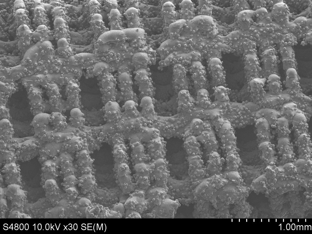
Grade 23 ELI Ti alloy
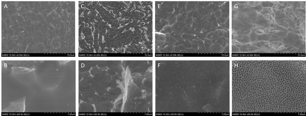
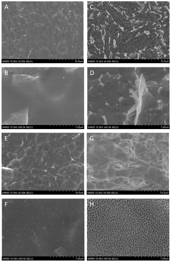
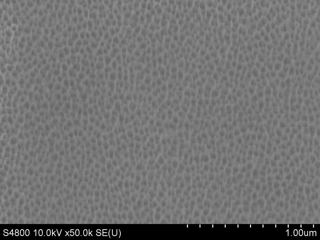
SEM images of Grade 23 ELI Ti alloy Shot Peened test surfaces at 5000x (A,C,E,G) and 50,000x magnification (B,D,F,H). ELI Ti (A,B); Nanorough acid etched (C,D); ELI Ti with CaP (E,F); Nanovis Bioceramic Nanotubes (G,H). CaP treatment present on (C,D,G,H).
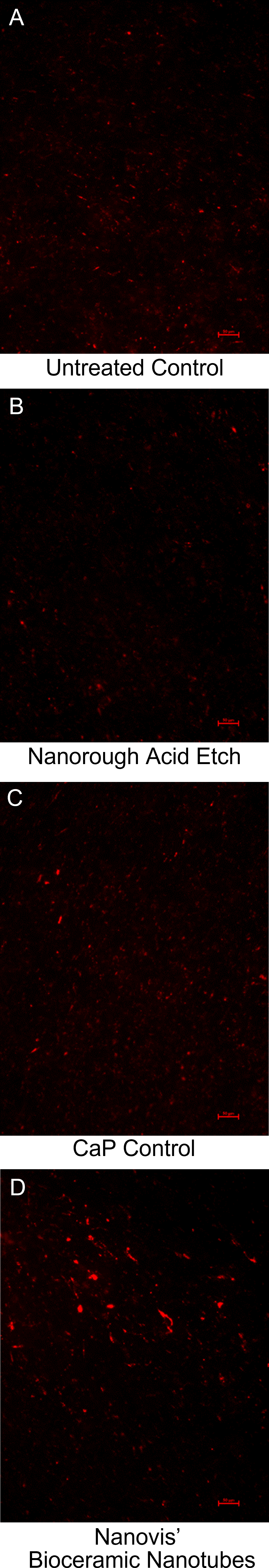
Osteoblast Mineralization
Day 21 (100x Magnification)
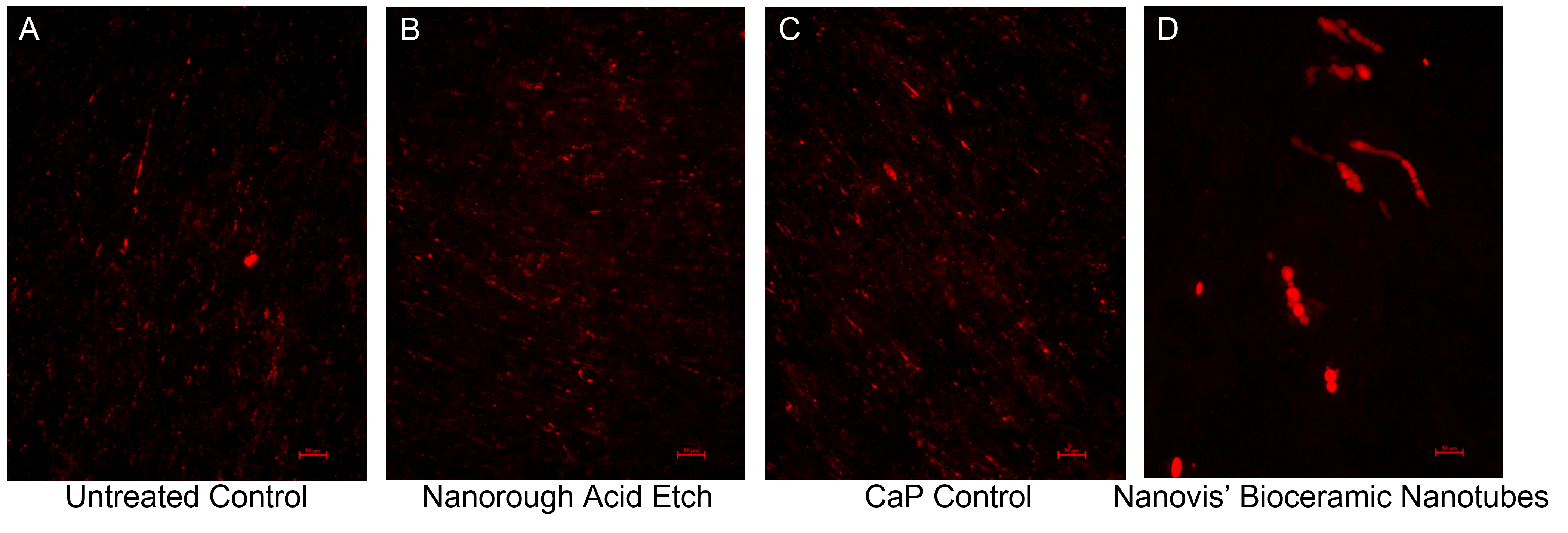
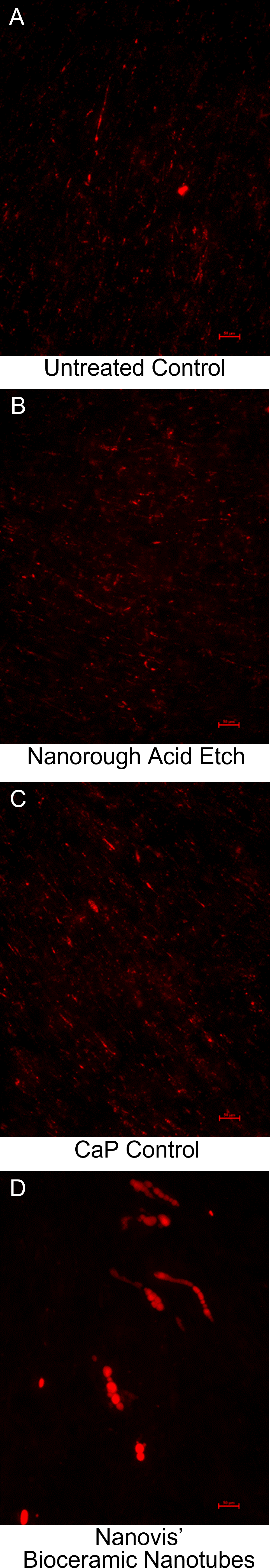
Mesenchymal Stem Cell Mineralization
Day 21 (100x Magnification)

Residual Bone
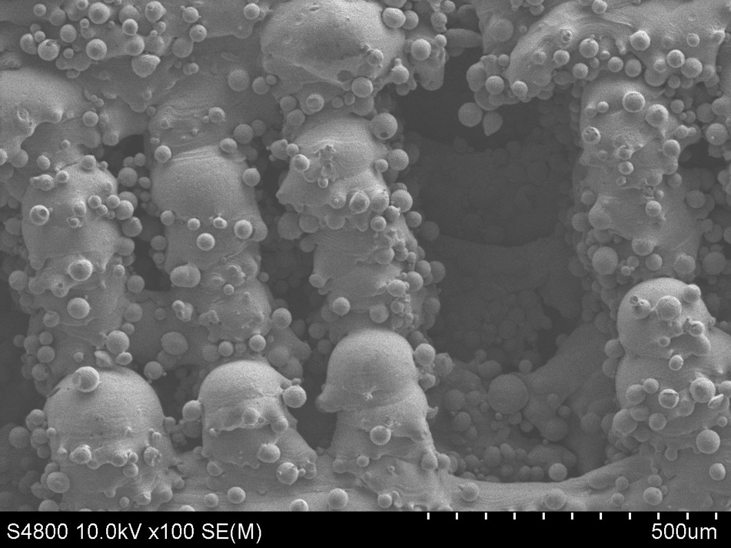
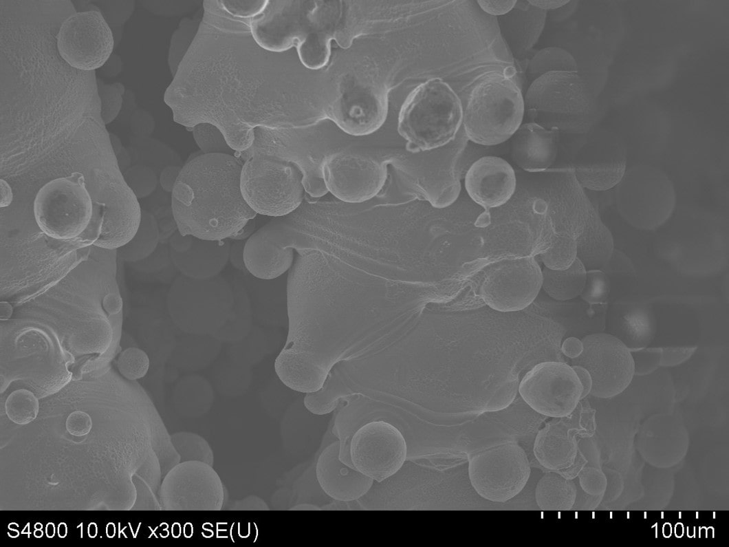
Flakes of residual bone on the surface of extracted pedicles screws in image A and B demonstrates that the bone failed while the bioceramic nanotubes remained attached to the screw. The bone matrix can be seen in image C and the collagen rich bone matrix can be seen attached to the nanotubes in D.
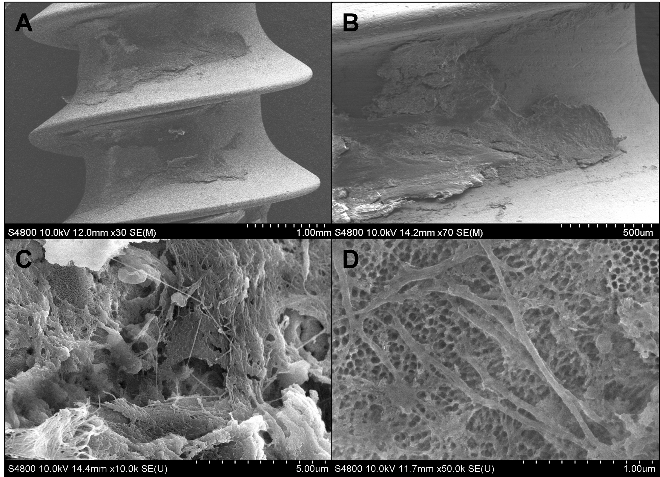
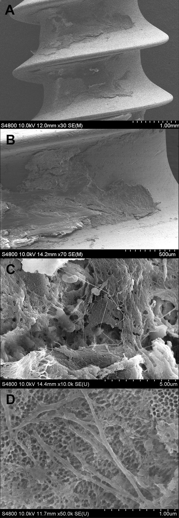
SEM images of residual bone on Nanovis’ Bioceramic nanotube pedicle screws after extraction from horse bone after 13 weeks of implantation. Magnifications: 30x (A), 70x (B), 10,000x (C), 50,000x (D).

Connect With Us
Columbia City, IN 46725
United States
Recent News